Total Pageviews

Sunday, 24 March 2013

Thoracic Outlet Syndrome due to a Cervical Rib

Cervical Rib Producing Thoracic Outlet Syndrome


A Cervical Rib is a rib arising from the 7th Cervical Vertebra. The charecteristic radiological feature of the a cervival vertebra is its transverse process which is directed downwards and laterally.
Whereas the transverse process of a Thoracic vertebra is directed upwards and laterally.This is the most important identification feature of a cervical rib in a plain radiograph.
The clincal manifestations of a cervical rib is called Thoracic Outlet Syndrome.
Thoracic outlet syndrome is neurovascular symptoms in the upper extremities due to pressure on the nerves and vessels in the thoracic outlet area.
What is meant by the Thoracic Outlet?
Thoracic Outlet is bounded
Anteriorly- Upper border of Manubrium Sterni
Laterally- First Rib on each side
Posteriorly- Superior srface of body of first thoracic vertebra

Antero posterior Diameter of the Thoracic Outlet is 5cm and the Transverse  Diameter 10cm.
The specific structures compressed are the Nerves of the Branchial plexus , Subclavian Artery or Subclavian Vein.
Aetiological Factors of Thoracic Outlet Syndrome
·Cervical Rib

·Congenital Fibrous Band Between First Rib & C7

·Abnormal Fusion of Scalene Muscles at the Insertion Site

·Clavicular Fracture Callus/ Traumatic

·Exostosis of First Rib/ Neoplastic

·Narrowing of Costo Clavicular Space

·Compression by Pectoralis minor Tendon

·Long Transverse process of C7

·Poor posture, ie Drooping Shoulders –Psychological     Depression  has to be ruled out.

·Repetitive activities, causing enlargement of the Scalenus    anterior muscle ( Lateral           Flexion     of Cervical Spine)
  Hence this type of Thoracic outlet syndrome is more common in Swimmers & Volley ball players.

·Post Sternotomy ( Pneumonectomy/ ASD Closure )

·Obesity/ Recent Weight Gain  causes – Narrowing of the Thoracic outlet.
 
Cervical Rib
·Arises from 7th Cervical vertebra
·Present in 0.5% of individuals
·May be Unilateral/ Bilateral
·Commoner on the Right Side
Types of Cervical Rib

1.     Complete Cervical Rib
2.     Cervical rib ending in a bony mass
3.     Cervical rib tapering as a Fibrous band ( Not visible in a Plain Radiograph )

4.     Complete Fibrous Band – No Radiological Finding

Clinical Features of Thoracic Outlet Syndrome
Upper Extremity
Arterial-  Numbness, Tingling & Weakness of Arms & Hands
Venous -  Swelling of Hand & Fingers, Pain
Neurological - Pain & Parasthesia over Ulnar / Median nerve distribution,Weakness & Coldness of the Hands, Tiredness& Heaviness on elevation of Arms.
Chest – Angina Like Pain/ Shoulder pain
Vertebral Artery Involvement- Dizziness/ Headache/Syncope
Note : In patients with Normal Coronary Angiogram & Normal Esophageal Function  having  Persistent Chest Pain Consider the possibility  of TOS
Clinical Signs of TOS
1.Difference in Pulse in both upperlimbs
2.Atrophy of  Thenar/Hypothenar  eminence
3.Brittle Nails / Focal Ulcers/Gangrene
4.The White Hand Sign –pallor of hand due to  Arterial TOS
5.Oedema of Hands – Venous TOS or Paget-Schroetter Syndrome or Effort Thrombosis.
Diagnostic Triad of Thoracic Outlet Syndrome or Selmonosky Triad 
1.     Tenderness in the Supraclavicular  Area
2.     Abduction & Adduction weakness of 4&5 fingers C8 - T1
3.     Pallor / Parasthesia on elevation of the Limb

Clinical Tests for Thoracic Outlet Syndrome
· Hyper abduction Manoeuvre- When arm is hyperabducted to 180°, neurovascular bundle components are compressed by the pectoralis minor tendon, leading to disappearance of radial pulse.

· Costoclavicular Compression   Manoeuvre- By keeping  the shoulders in backward and downward position as in exaggerated military position, causes reduction in costo clavicular space  and compression of subclavian artery leading to disappearance of radial pulse.


· The Elevated Arm Test or  Roose Test- Arm abducted at 90 degree with external rotation of the shoulder. Opening & closing of hands done rapidly for 3 minutes. A normal individual can do this  without any difficulty. In TOS patient develops pain and parasthesia of fore arm muscles and fingers.


Clinical Investigations for Thoracic Outlet Syndrome

1. Plain Radiograph of Neck

2.MRI Scan

3.Nerve Conduction Velocity Study


Differential Diagnosis of Thoracic Outlet Syndrome

1.     Cervical Spondylosis

2.     Cervical Disc Prolapse

3.     Cervical Cord Compression

4.     Carpel Tunnel Syndrome

5.     Raynaud’s disease / Pancoast Tumour of Lung


Treatment of Thoracic Outlet Syndrome- Conservative
1.     Maintain a Proper posture

2.     Weight Reduction ( Obesity predisposes to TOS )

3.     Physiotherapy- Exercise to strengthen the  Shoulder girdle to prevent drooping.

4.     Stop any repetitive activity or work for prolonged period


Indications of Surgical Intervention in Thoracic Outlet Syndrome

1.     Failure of Conservative Management for 6 months

2.     Progression of the Neurological Symptoms

3.     Occlusion of Subclavian Artery

4.     Thrombosis of Subclavian/ Axilliary vein


Surgical Excision of the Cervical Rib or Scalenotomy Operation
— 
Anterior Approach

Posterior Approach

Trans Axillary Approach
    
Important Operative Surgery Steps
Lateral Supra Clavicular Incision

Platysma& Deep fascia incised

Sternomastoid  retracted medially

Phrenic nerve is identified over the Scalenus anterior muscle

Scalenus anterior muscle is divided at its insertion to first rib after safeguarding the Phrenic nerve

Cervical rib is divided along with the Periosteum to prevent recurrence.
  
Complications of the Surgery for Cervical Rib
1.     Phrenic nerve injury

2.     Injury to Brachial plexus

3.     Injury to Subclavian artery and vein

4.     Pneumothorax


Thursday, 21 March 2013

What is a T Tube Cholangiogram?

 T- Tube Cholangiogram

What is a T Tube Cholangiogram?
Presence of stone inside the common bile duct is called Choledocholithiasis.
There are two ways of stone formation occurring in side the common bile duct. One is from the gall bladder.
The other is de novo stone formation inside the common bile duct.
The cause of de novo stone formation inside the common bile duct is Portal pyemia  or cholangitis.
The stone formed as a result of portal pyemia is Brown pigment stone. Black pigment stone is formed as result of excessive haemolysis.
After the open surgical exploration for Common bile duct for choledocho lithiasis, the completion of the stone removal is not full proof, as the stone removal is a blind process.
 That means, in open surgery, even after the removal of last stone from the common bile duct one is not sure that all the stones are out.
Hence a special tube called T tube is placed. The upper horizontal part of the T tube is placed inside the common hepatic duct.
The lower horizontal part of the T tube is placed inside the distal common bile duct. The vertical part of the T tube is brought out to skin surface.
If there is a distal obstruction of the common bile duct the quantity of bile coming out through the vertical limb will be more.
The normal amount of bile secreted by the liver is around 800-1000ml. Hence if the discharge through  the T tube is approaching this amount it is presumed that there is distal obstruction for the bile flow.
To confirm the distal obstruction on 10th post operative day we do an
investigation called T tube cholangiogram.
In the T tube cholangiogram if the radio opaque iodine containing dye is reaching the duodenum that means there is no distal obstruction.
The left over stones are seen as the filling defects or negative shadows.
The special forceps used to remove the stones from the common bile duct is called Desjardin’s Choledocholithotomy forceps.
The dilators we use for dilating the common bile duct is called Bake’s Dilators. These are dilators with serially increasing diameters.
The latest device used in the removal of stones from the common bile duct is called Choledochoscope.
Through a Choledochoscope one can see the interior of the common bile duct as well as the common hepatic duct  and all the stones can be removed under vision, making a T tube cholangiogram  not mandatory.


Tuesday, 12 March 2013

What is a Sebaceous Cyst?

What is a Sebaceous Cyst?
A sebaceous cyst is a retention cyst arising from a Pilo sebaceous Gland.


Clinical Features of  Sebaceous Cyst:-

  • Skin not pinchable
  • Presence of a Punctum
  • Indentation possible due to pultaceous material.
  • Not fixed to underlying Structures.

Multiple Sebaceous Cysts are called -Wen

Common sites of Multiple Sebaceous Cysts- Scrotum & Scalp

Complications of  Sebaceous Cyst:-

  • Infection
  • Ulcerations
  • Calcifications
  • Sebaceous Horn
  • Malignant Transformation ( Rare)- Squamous Cell Carcinoma

A Parasite found in a Sebaceous Cyst :- Demodex follicularum

Treatment of  Sebaceous Cyst:- 
Excision of the Sebaceous Cyst.

What is the Incision for Excision of the Sebaceous Cyst?
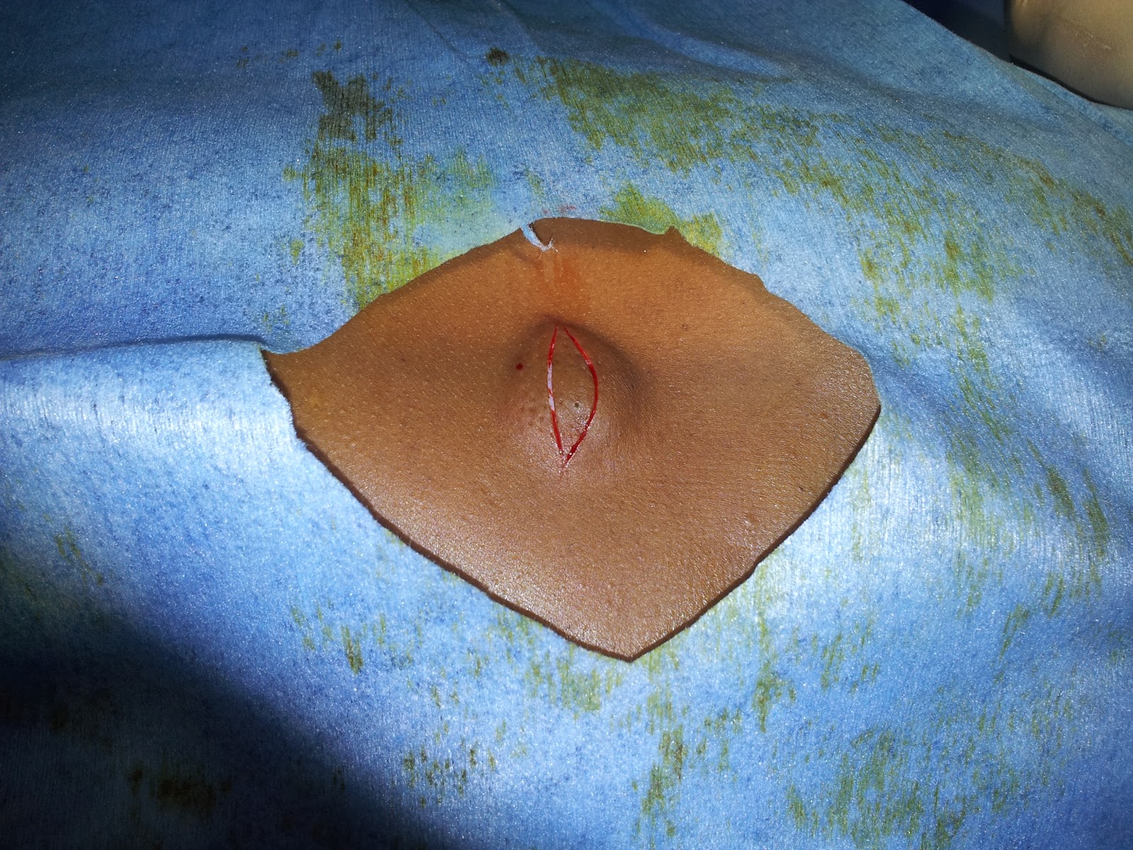
Elliptical Incison encircling the Punctum of the Sebaceous Cyst.

What is the correct name of the Incision for Excision of the Sebaceous Cyst?
The correct name of the Incision is - Lentiform ( Biconvex )Incision.

A sebaceous cyst cannot occur in Palm and Sole as there is no Sebaceous Gland in these sites.

Saturday, 9 March 2013

Multiple Perforator Ligations for Varicose veins

Multiple Perforator Ligations for Varicose veins due to Perforator Incompetence.


   The open surgery for varicose veins due to Perforator Incompetence is identification and ligation of the marked perforators . Note the T junction in the course of the Great Saphenous Vein.
    The perforators pierce the deep fascia of the lower limb and connects with the deep venous system. This typical T junctions are ligated at 3 points, that is, two horizontal points and one vertical point. Hence this surgery is also called Multiple ligations.

Wednesday, 6 March 2013

Surgery For Hydrocele- Jaboulay’s Procedure or Eversion TV Sac


Surgery For Hydrocele- Jaboulay’s  Procedure or Eversion TV Sac


Incision:
Vertical Paramedian ( Note the Hyperpigmented Median Raphae- -in Figure 1)
Procedure-
Step 1. Incision deepened and the Hydrocele Sac Isolated

Step 2. Hydrocele Fluid  drained and Excess sac excised.

Step 3. Eversion of TV Sac ( Tunica Vaginalis sac).

Step 4. Wound is closed in layers after achieving complete hemostasis.


Step 5. Scrotal suspensory bandage applied ( Scrotal Support )


Saturday, 2 March 2013

Sub Platysmal Flap for Open Thyroidectomy

The Flap Raised for Open Thyroidectomy is called Sub Platysmal Flap.


It is raised in a plane after cutting the subcutaneous muscle, Platysma.
The superior extent of the flap is up to the level of Thyroid cartilage and the inferior extend is up to the Supra sterna notch.

Charecteristic Features of Arterial Claudication Pain


Charecteristic Features of Arterial Claudication Pain


Arterial Claudication Pain is due to the Ischemia to Muscles.

1.     Claudication Pain is always precipitated by walking/exercise.
2.     Claudication Pain is relieved by stoppage of walking/exercise.
3.     Claudication Pain is always felt over Muscles.
4.     Claudication Pain is always reproducible.