Total Pageviews

Sunday 23 June 2013

Mesenteric Cyst

Mesenteric Cyst


Mesenteric Cyst

                 
 Mesenteric Cysts are cysts arising from the mesentery.
Types of Mesenteric cysts:-
1.     Chylo lymphatic cyst ( Commonest )
2.     Enterogenous cyst
3.     Urogenital Remnant
4.     Teratomatous Dermoid Cyst (Least common)
Commonest age group- Second decades of Life
What is the characteristic clinical feature of a Mesenteric Cyst?
Painless cystic intra abdominal swelling in the umbilical region which moves freely in a plane perpendicular to the line of attachment of root of mesentery.
Differential Diagnosis
Omental cyst/ Ovarian cyst
Complications
  • Torsion
  • Rupture
  • Infection
  • Hemorrhage into the cyst
What is the difference in Blood supply of a Chylo lymphatic cyst and an Enterogenous cyst?
Blood supply of a Chylo lymphatic cyst is independent of the adjacent intestine.Hence simple excision is possible.
But Enterogenous cyst derives its blood supply in common with the adjacent potion of the intestine.
Hence surgical removal of an enterogenous cyst entails the resection of the related segment of the intestine.
                        

Basic Principles of Laparoscopic Surgery-Part 2

Basic Principles of Laparoscopic Surgery-Part 2
 
Laparoscopic surgery is an instrument based surgery. The success of a laparoscopic procedure depends on the skill of the surgeon and the combined efforts of his surgical team.
Co axial / straight line principle of Laparoscopy:
The surgeon, the target organ of pathology and the monitor must be in a straight line.
Base Ball Diamond Court Concept of Laparoscopy:


                                        Pathology


 Right Port                                                            Left Port
                                      

                                      Centre Port

                                             
Baseball Diamond Court Concept is important in the triangulation of the instruments. If this principle is followed the clashing between the instruments can be prevented.

Manipulation Angle is the angle between two working ports. Ideal manipulation angle is 60º.
Azimuth Angle is the angle between one instrument and the optical axis of the endoscope. Ideal azimuth angle is equal for the active and assisting instruments.
Elevation Angle is the angle between the instrument and the horizontal plane. Ideal elevation angle is 60º.
These three angles determine the optimal port placement.


Saturday 22 June 2013

Bilateral Carcinoma Breast

Bilateral Carcinoma Breast


When the patient presents with features of Bilateral Breast Cancer, there are two possibilities.
The contralateral cancer can be a Second primary cancer or a Metastatic lesion.
The differentiation is very important in the management.
Synchronous Primary Cancer-when the two primary cancers develop with in 6 months
Metachronous Primary Cancer- when the second cancer develop after 6 months.
Histological grade, Immunohistochemistry, ERPR Status of both lesions must be studied. These are similar for a metastatic lesion and are different for a second primary lesion.
A complete metastatic work up for the patient is also necessary to rule out hepatic, lung, bone or brain metastasis.
If there is no evidence of metastasis, the diagnosis goes in favor of the second primary lesion.
Chaudary’s criteria is widely followed for the diagnosis of a second primary breast cancer.
Chaudary’s criteria
1.There must be insitu changes in the contralateral tumour.
2.The second tumour is histologically different from the first.
3.The histological differentiation grade of the second tumour greater than the first.
4.There must be no evidence of  metastasis.

Thursday 20 June 2013

Branchial Cyst

Branchial Cyst Excision

Branchial Cyst Excision


Branchial Cyst Excision


Branchial Cyst

Common Age Group- First or second decades of life
Site- Along the Anterior border of the Sterno Mastoid at the junction between Upper one third and lower two third.
Shape-Spherical or Ovoid
Surface-Smooth
Consistency-Soft
Transillumination test- Positive if it contains clear fluid
Mobility- Restricted mobility
Plane of the Swelling- Deep to Sterno Mastoid Muscle
Compressibility- Negative
Aspiration fluid contains-Cholesterol crystals

Differential Diagnosis:
  • Lymph Nodular Enlargement
  • Dermoid Cyst
  • Vascular/ Lymphatic Malformations

Complication: Infection
Treatment: Complete Surgical Excision under General Anesthesia.
(Incomplete excision can result in recurrence.)
Cystic swelling containing Cholesterol Crystals in the aspiration fluid:-
  1. Branchial Cyst
  2. Thyroglossal cyst
  3. Hydrocele
  4. Cystic Hygroma
  5. Detigerous Cyst (- arises from an Un erupted tooth )
  6. Dental Cyst (- is a complication of Dental caries )

Tuesday 18 June 2013

Gastro Intestinal Fistula

Gastro Intestinal Fistula
Definition
A fistula is tract connecting two epithelial surfaces one of which is a hollow organ.
Classifiaction of Fistulae
Internal Fistula:-
A fistula connecting any two digestive organs.
External Fistula:-
A fistula connecting  a digestive organ and skin.
Uncontrolled fistula:-
A fistula which contaminates the general peritoneal cavity is called  uncontrolled fistula.
Controlled fistula
If the effluent of a fistula is drained out using a Foley’s catheter, without contaminating the general peritoneal cavity, it is called  controlled fistula.

Based on the out put fistulae can be
Low output Fistula :– If the output is less than 200 ml/24 Hours
Moderate output Fistula:– If the output is between 200-500ml/24 Hours
High output Fistula:– If the output is more than 500ml/24 Hours


Causes of Gastro Intestinal Fistula

  • Congenital
  • Traumatic
  • Inflammatory-Cohn’s Disease
  • Neoplastic-Carcinoma colon
  • Post operative-Post appendicectomy /Intestinal Anastomosis.

Complications of GI Fistula
  • Sepsis
  • Fluid and Electrolyte Imbalance
  • Malnutrition
  • Effluent Dermatitis-resulting in skin excoriations, ulcerations and infections.

Principles of Management of GI Fistula:-


  • Correction of Fluid and Electrolyte Imbalance.
  • Nil Per Oral and Bowel rest.
  • Total Parenteral Nutrition.
  • Conversion of an uncontrolled fistula into a controlled fistula.

Prevention of Effluent Dermatitis:-
By painting the skin with Zinc oxide paste.
MCQ
The commonest electrolyte abnormality associated with a GI Fistula is Hypokalemia.

Port Site Infection

 The Port Sites


Port Site Infection
The Commonest Organism Causing Port Site Infection Following Laparoscopic Procedures is Atypical Mycobacteria.
Hence the treatment recommended for Port Site Infection is Anti Tuberculosis Treatment.
How to prevent the Port Site Infection?
Port Site Infection can be prevented by following strict sterilization of the laparoscopic instruments.  

Port Site Hernia
Port site hernia is the hernia occurring through the large port sites like 10mm Ports.
How to prevent Port Site Hernia?
Port Site Hernia is prevented by proper closure of the Anterior Rectus or the Linea Alba with Poly propylene suture material.

Port Site Recurrence
Port Site Recurrence of malignancy after a laparoscopic procedure can occur if adequate precautions are not taken.
How to prevent Port Site Recurrence?
Port Site Recurrence can be avoided by using an ‘Endo Bag’ for the retrieval of the specimen.


The Cause of Shoulder pain following Laparoscopy

What is the cause of shoulder pain following laparoscopic procedures?

The commonest distension medium used in laparoscopy is carbon dioxide gas. Carbon dioxide is 20 times more soluble in body fluids than air or oxygen.
When pneumoperitoneum is created with carbon dioxide during a laparoscopic procedure, Carbonic acid is formed inside the abdomen.
Carbonic acid becomes adherent to diaphragm . The diaphragmatic irritation with  Carbonic acid  is responsible for the shoulder pain following Laparoscopic procedures.
The ideal solution to prevent this adhesion of carbonic acid to the diaphragm  is  Ringer lactate solution.

How much peritoneal wash should be  given with Ringer lactate?
Peritoneal wash must be given with warm  Ringer lactate till the crystal clear solution stays inside the abdomen.
Some amount of Ringer lactate solution has to be left inside the abdominal cavity for the lubrication of the intestines, thus  preventing future adhesions.
( Normally also there is some amount of biologic fluid inside the abdomen for lubrication. Hence it is not recommended to suck out all the irrigation fluid back from the peritoneal cavity after an abdominal surgery)

Sunday 16 June 2013

Basic Principles of Laparoscopic Surgery - Part 1

Basic Training in Laparoscopic Surgery


Basic Training in Laparoscopic Surgery includes:-
  • Understanding the fundamental principles of laparoscopy
  • Mastering the art of Instrumentation
  • Hands on Training
  • Supervised practice on humans (Most important)
  • To know one’s role as a Team work(Very important)

Fundamental principles of laparoscopy:-
Pneumoperitoneum is created using a distention medium like Carbon dioxide with the help of an Insufflator machine.
Since carbon dioxide is soluble in blood a capnograph is a must in monitoring the patient during anesthesia for laparoscopic surgeries.
Safe method of insufflation is by using a spring loaded Veress needle.
Insufflation is completed by the introduction of Primary Trocar and the Telescope. This is called the camera port.
The telescope is in turn connected to a light source, fiber optic cable and to a video monitor.
Other ancillary instruments like forceps, scissors, electro cautery, needle holder, clip applicator, suction irrigation device etc are also used. Almost all instruments used in open surgery are now available for laparoscopic surgery.
These instruments are introduced through different ports. The positions of primary port and the accessory ports are determined by the principles of Ergonomics for laparoscopic surgery. (Ergonomics means the norms of work)
Hands on training is a must for the proper hand eye coordination. Good coordination between right and left hands are also needed for laparoscopic procedures.

Supervised practice on humans is another important aspect of training. Each member of the team should know his or her role  for a particular procedure.

Saturday 8 June 2013

Obesity, Morbid Obesity, Super Obesity, Super Super Morbid Obesity

Obesity, Morbid Obesity, Super Obesity, Super Super Morbid Obesity:-

Bariatric Surgery for Morbid Obesity
Body Mass Index is the ratio of Body weight in Kg and the square of the Height in metres.

Normal Body Mass Index (BMI) 18.5-24.9 Kg/m2
Overweight is Body Mass Index  25-29.9 Kg/m2
Obesity is defined as a Body Mass Index > 30 Kg/m2
Morbid obesity is Body Mass Index > 40 Kg/m2
Super obesity is Body Mass Index > 50 Kg/m2
Super super obesity (body mass index (BMI)>60 Kg/m2

Bariatric Surgery means surgery done for the correction of Morbid Obesity, Super Obesity, Super Super Morbid Obesity.

Two most popular techniques of  Bariatric Surgery are:-
1.Laparoscopic Sleeve Gastrectomy ( LSG )
2.Roux-en-Y Gastric Bypass ( RYGB )

Bariatric Surgery surprisingly induces remission of the metabolic disorders associated with obesity like Hypertension, Diabetes, Obstructive Sleep Apnoea and Dislipidemia.

Hence if such bariatric surgical procedure is done for the correction of a metabolic condition like Diabetes mellitus with failed medical therapy in a non-obese person  is termed as Metabolic Surgery.


Foreign Body Inside the Urinary Bladder

Foreign body inside the Urinary Bladder- A Case Report
Wooden Foreign body inside the Urinary Bladder

Case Capsule
70 year old male patient, presented to the emergency surgical  department with history of accidental fall  over a wooden piece .
 With complaints of pain in the lower abdomen and wound over the peri anal region.
 On admission patient was hemodynamicaly stable.
He did not pass urine since the event.
No associated  bleeding per meatus or fecal incontinence
Per Abdomen: showed diffused lower abdominal tenderness.
 No rigidity noted.
Bladder not palpable per abdomen.
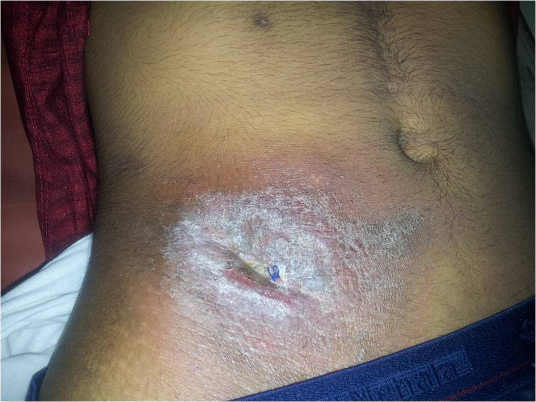
Local Examination
  Lacerated wound over the perianal region at 7’o clock position measuring approx 4 x 2 cm extending to the anterior rectal wall.
Digital Rectal Examination
 Lax sphincter tone
Approx 5 cm linear defect palpated over the anterior rectal wall starting approx about 2cm from the anal verge, communicating with external perianal wound.
With fecal contamination of the perianal wound noted.
CT scan taken revealed :
Rectovesical fistula tract extending from the perianal external wound to the base of the bladder.
Linear tear involving the anterior rectal wall.
Tear involving the base of the bladder.
Foreign body inside the bladder.
Urology consulatation was taken and planned for cystostomy and evacuation of the vesical foreign body.
Patient was posted for the emergency surgery.
Intra op findings and the Procedure
A linear tear of the anterior rectal wall was noticed starting approximately  2cm from the anal verge.
 With a full thickness rectal rent noted in its most proximal portion of the tear just below the peritoneal reflection communicating with the base of the bladder.
Tear involving the base of the bladder measuring approx 1 x 0.5 cm just above the inter ureteric ridge noted.
Wooden piece noted with in the bladder cavity, which was extracted.
Bilateral ureteric orifice normal.
Bilateral  DJ stenting done.
Vesical tear closure done.
SPC  done.
Vertical cystostomy closure done.
Rectum mobilised and primary repair  of the rectal tear done.
Diversion clostomy done via sigmoid loop colostomy.
Perianal wound was kept open.
Post Operative Period 
Post op period was uneventful.
Was started on enteral feeds on the post operative day 2.
Was discharged onpost operative day 12 with insitu spc and uretheral catheter.
                                        Courtesy : Dr Mohammed Jashin