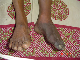
Clinical Examination of an Ulcer
Definition of an Ulcer
— An ulcer is defined as a break in the continuity of surface epithelium with superadded infection.
Clinical Classification of Ulcer
— Spreading- with surrounding inflammation
— Healing – Slopping edge with red granulation tissue
— Callous- Ulcer with no tendency to heal-with pale granulation tissue.
Pathological Classification of Ulcer
— Specific- Tuberculous, Syphylitic,Actinomycotic
— Non specific- Traumatic( Mechanical, Physical,Chemical)
Cryopathic, Arterial, Venous, Neurogenic, Trophic, Tropical, Bazin’s, Martorell’s, Meleney’s ulcer
— Malignant- Squamous cell carcinoma, Basal Cell Carcinoma, Melanoma
Classification of Ulcer based on duration
— Acute Ulcers < 12 weeks
— Chronic Ulcers > 12 weeks
Classification Based on Pain
— Painful Ulcers
Tuberculous
Arterial
Advanced Malignancy
— Painless Ulcers-
Syphilitic
Trophic
Early Malignancy
Modes of Onset of Ulcer
• Traumatic
• Spontaneous-
• Secondary changes on a Swelling-Tuberculous lymphadenopathy
• From a Previous Scar-Marjolin’s Ulcer
Causes for Chronic Ulcers
1. Malnutrition
2. Anemia
3. Immunosuppression
4. Systemic Diseases (Diabetes)
5. Site & Size of an Ulcer
6. Arterial / Venous Disorders
7. Neurological Disorders
8. Infection
9. Chronic Irritation ( Dental Ulcers ) or Lack of Rest ( Ulcer over a Joint )
10. Malignancy
Discharge from the ulcer – Note the Colour, Amount & Smell
— Serous-Healing ulcer
— Sanguinous ( Blood Stained )- Malignant/Chronic
— Purulent-Bacterial infection
— Greenish- Pseudomonas infection
— Yellowish - Sulphur Granules ( Actinomycosis )
Parts of an Ulcer
• Margin-line of demarcation between normal and abnormal
• Floor-the exposed part of an ulcer ( Inspection)
• Edge-the part between the margin and the floor of an ulcer
• Base-the structure on which the ulcer rests (Palpation)
Types of Floor of an Ulcer
— Slough-Moist dead tissue
— Scab-Dry dead tissue
— Unhealthy granulation tissue
— Healthy granulation tissue
— Subcutaneous fat
— Muscle/tendons
— Bone
Types of Edges of an Ulcer
— Slopping- Healing ulcer
— Punched out-Decubitous ulcer/Gummatous ulcer
— Undermined- Tuberculous ulcer
— Raised / Beaded- Basal cell carcinoma
— Rolled out / Everted- Squamous cell carcinoma
Examination of an Ulcer-Inspection
— Site
— Size
— Shape
— Number
— Margin
— Edge
— Discharge
Importance of Site of Ulcer
Face-Basal Cell Carcinoma
Neck-Tuberculous/Actinomycotic
Decubitus ulcer-Over pressure points like sacral/occiput/heel
Shin-Gummatous
Medial malleolus-Varicose ulcer
Examination of an Ulcer – Palpation
— Local rise of Temp & tenderness
— Exact dimensions - depth
— Induration ( thickening ) of edge-in chronic ulcer and in malignancy
— Base – fixity to underlying structures
— Bleeding on touch-is a feature of malignant/ Chronic ulcer
Examination of the Surrounding Area of an Ulcer
— Skin
— Adjacent Joint
— Regional Lymph nodes
— Arterial pulse
— Varicose veins
— Neurological deficit
— Gait of the patient
A Golden Rule
Whenever you see a lesion look for an enlarged draining lymph node;
Whenever you come across an enlarged lymph node in our body look for a lesion in the area of its lymphatic drainage.


